| Size | Price | Stock | Qty |
|---|---|---|---|
| 5mg |
|
||
| 10mg |
|
||
| 25mg |
|
||
| 50mg |
|
||
| 100mg |
|
||
| 250mg |
|
||
| 500mg |
|
||
| Other Sizes |
|
Purity: ≥98%
GSK690693, an aminofurazan derivative, is a novel, potent and ATP-competitive pan-Akt inhibitor targeting Akt1/2/3 with potential anticancer activity. With IC50 values of 2 nM, 13 nM, and 9 nM in cell-free assays, it inhibits Akt1/2/3. GSK690693's antiproliferative effects were specific to cancer cells because it had no effect on the growth of healthy mouse thymocytes or normal human CD4(+) peripheral T lymphocytes. GSK690693 treatment decreased the phosphorylation of downstream AKT substrates in both sensitive and insensitive cell lines, indicating that the lack of AKT kinase inhibition was not the root cause of resistance.
| Targets |
Akt1 (IC50 = 2 nM); Akt3 (IC50 = 9 nM); Akt2 (IC50 = 13 nM); PKCη (IC50 = 2 nM); PKCθ (IC50 = 2 nM); PrkX (IC50 = 5 nM); PAK6 (IC50 = 6 nM); PAK4 (IC50 = 10 nM); PKCδ (IC50 = 14 nM); PKCβ1 (IC50 = 19 nM); PKCε (IC50 = 21 nM); PKA (IC50 = 24 nM); PKG1β (IC50 = 33 nM); AMPK (IC50 = 50 nM); PAK5 (IC50 = 52 nM); DAPK3 (IC50 = 81 nM); Autophagy
GSK-690693 inhibits Akt1 (IC50 = 2 nmol/L, Ki = 1 nmol/L); Akt2 (IC50 = 13 nmol/L, Ki = 4 nmol/L); Akt3 (IC50 = 9 nmol/L, Ki = 12 nmol/L); additionally inhibits other AGC kinase family members including PKA (IC50 <100 nmol/L), PrkX (IC50 <100 nmol/L), PKC isozymes (IC50 <100 nmol/L), AMPK (IC50 <100 nmol/L), DAPK3 (IC50 <100 nmol/L), and PAK4/5/6 (IC50 <100 nmol/L). |
|---|---|
| ln Vitro |
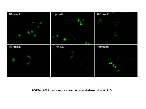
GSK690693 is very selective for the Akt isoforms versus the majority of kinases in other families. With IC50 values of 24 nM, 5 nM, and 2-21 nM, respectively, GSK690693 is less selective for AGC kinase family members such as PKA, PrkX, and PKC isozymes. Additionally, PAK4, 5, and 6 from the STE family have IC50 values of 10 nM, 52 nM, and 6 nM, respectively. GSK690693 also potently inhibits AMPK and DAPK3 from the CAMK family. With an IC50 ranging from 43 nM to 150 nM, GSK690693 prevents tumor cells from phosphorylating GSK3. The nuclear accumulation of the transcription factor FOXO3A is increased dose-dependently by treatment with GSK690693. The IC50 values for GSK690693 are as follows: 72 nM, 79 nM, 86 nM, 119 nM, 975 nM, and 147 nM for T47D, ZR-75-1, BT474, HCC1954, MDA-MB-453, and LNCaP cells, respectively. In both LNCaP and BT474 cells, treatment with GSK690693 induces apoptosis at concentrations >100 nM. [1] GSK690693 induces apoptosis in susceptible ALL cell lines, supporting the function of AKT in cell survival.[2]
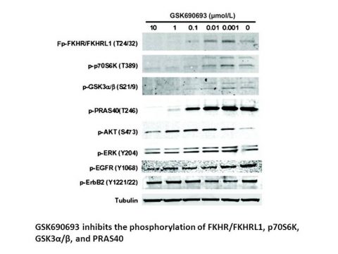
GSK-690693 inhibited phosphorylation of GSK3β (Ser9) in tumor cells with average IC50 values ranging from 43 to 150 nmol/L across multiple cell lines (e.g., LNCaP: 43 nmol/L, BT474: 160 nmol/L, 786-0: 150 nmol/L), as measured by ELISA. In BT474 breast cancer cells, GSK-690693 induced dose-dependent reductions in phosphorylation of Akt substrates including FKHR/FKHRL1 (Thr24/32), p70S6K (Thr389), GSK3α/β (Ser21/9), and PRAS40 (Thr246) at concentrations >100 nmol/L, confirmed by Western blot. Phosphorylation of Akt (Ser473) increased at low concentrations (1 nmol/L–1 μmol/L) but decreased at 10 μmol/L. GSK-690693 suppressed proliferation in sensitive tumor cell lines: IC50 values were 86 nmol/L (BT474), 147 nmol/L (LNCaP), 119 nmol/L (HCC1954), and 72–79 nmol/L (T47D, ZR-75-1). Insensitive lines (e.g., MDA-MB-231, HT29) had IC50 >10 μmol/L. Proliferation was measured via CellTiter Glo assay after 72-hour treatment. In FOXO3A-GFP translocation assays, GSK-690693 (≥1 μmol/L) induced nuclear accumulation of FOXO3A in U2OS cells within 90 minutes, indicating functional inhibition of Akt-mediated phosphorylation. GSK-690693 induced apoptosis in LNCaP and BT474 cells at concentrations >100 nmol/L after 24–48 hours, quantified by histone-complexed DNA fragments. [1] GSK690693 inhibited proliferation in 55% (62/112) of hematologic malignancy cell lines (EC50 < 1 µM). Acute lymphoblastic leukemia (ALL) showed highest sensitivity (89%, 31/35 cell lines), including 95% T-ALL (19/20) and 80% B-ALL (12/15). Non-Hodgkin lymphoma (73%) and Burkitt lymphoma (67%) were also highly sensitive, while AML (18%), CML (0%), and Hodgkin lymphoma (25%) were less responsive. In sensitive ALL cell lines (e.g., A3, I2.1, I9.2), GSK690693 induced dose-dependent apoptosis, evidenced by: - Increased sub-2n DNA content (cell death) and reduced S-phase cells. - Cleaved caspase-3 elevation (24 hours). - PARP cleavage and caspase 3/7 activation. - DNA fragmentation (72 hours, Cell Death ELISA). GSK690693 suppressed phosphorylation of AKT substrates (GSK3β-Ser9, PRAS40-Thr246, p70S6K-Thr421/Ser424) in both sensitive (A3) and insensitive (Tanoue) ALL cell lines at 6 hours (Western blot). Feedback hyperphosphorylation of AKT (Ser473/Thr308) occurred at low concentrations. **Selectivity for Malignant Cells**: Normal human CD4+ T cells (EC50 > 10 µM) and mouse thymocytes (EC50 > 30 µM) were unaffected, contrasting with PI3K inhibitor ZSTK474 (EC50 < 1 µM in normal lymphocytes).[2] Primary Thymic Lymphoma Cells: GSK690693 reduced viability in tumor cells from Lck-MyrAkt2 mice (IC50 ≈ 0.3–5 μM, MTT assay). Sensitivity varied across isolates (e.g., 55-1143 IC50 ≈ 0.3 μM vs. 55-228 IC50 ≈ 5 μM after 72 hrs). Ovarian Carcinoma Cells: Differential sensitivity observed: MOVCAR5 cells (IC50 ≈ 3 μM) responded to GSK690693, while MOVCAR6 cells were resistant. Human SKOV3 cells (IC50 ≈ 3 μM) served as positive control. Apoptosis & Cell Cycle: - Thymic lymphoma cells showed 2–3-fold apoptosis increase (annexin V/PI staining) after 24-hr treatment (10–20 μM). - MOVCAR5 cells exhibited ~50% G1-phase arrest (no significant apoptosis). Western Blot: 10 μM GSK690693 downregulated p-GSK3α/β, p-mTOR, p-p70S6K, and p-Akt1s1 (PRAS40) in thymic lymphoma and ovarian cells within 8–24 hrs. Feedback p-Akt upregulation was consistently observed. [3] |
| ln Vivo |
GSK690693 inhibits GSK3 phosphorylation in human breast carcinoma (BT474) xenografts in a dose- and time-dependent manner after a single administration. The Akt substrates PRAS40 and FKHR/FKHRL1 are reduced in phosphorylation when GSK690693 is administered. Blood glucose levels are also acutely elevated by GSK690693 but return to normal 8 to 10 hours later. The growth of human SKOV-3 ovarian, LNCaP prostate, and BT474 and HCC-1954 breast carcinoma xenografts is potently inhibited by the administration of GSK690693, with a maximal inhibition of 58% to 75% at the dose of 30 mg/kg/day.[1] GSK690693 is effective regardless of the Akt activation mechanism at play. When administered to Lck-MyrAkt2 mice, which express a membrane-bound, constitutively active form of Akt, GSK690693 slows the progression of tumors the best. [3]
In BT474 breast carcinoma xenografts (SCID mice), a single intraperitoneal (i.p.) dose of GSK-690693 (10–40 mg/kg) inhibited GSK3β phosphorylation dose-dependently (up to 87% inhibition at 40 mg/kg). Tumor drug concentrations >3 μmol/L (∼1,500 ng/g) correlated with sustained ∼60% reduction in phosphorylation for 8 hours. Daily i.p. administration of GSK-690693 (10–30 mg/kg for 21 days) significantly inhibited tumor growth in xenograft models: 58–75% inhibition in SKOV-3 ovarian, LNCaP prostate, BT474 breast, and HCC-1954 breast carcinomas (P < 0.01–0.05 vs. vehicle). Immunohistochemistry (IHC) of BT474 xenografts after repeat dosing showed reduced phosphorylation of Akt substrates PRAS40 (Thr246) and FKHR/FKHRL1 (Thr24/32), confirming in vivo target modulation. Acute hyperglycemia and increased plasma insulin (peak 643 ng/mL at 4 hours vs. baseline 0.73 ng/mL) occurred post-dosing, resolving by 24 hours. This metabolic effect aligned with circulating drug kinetics and was reversible. [1] Lck-MyrAkt2 Thymic Lymphoma Model: - GSK690693 (30 mg/kg i.p., 5 days/wk × 4 wks) reduced tumor incidence: 52% tumors in treated vs. 90% in placebo. - Tumor volume decreased 2-fold (112 mm³ vs. 231 mm³, p=0.026). - IHC confirmed ↓Ki-67 (proliferation; p=0.001), ↑cleaved caspase-3 (apoptosis), and ↓p-FoxO1/3 cytoplasmic staining. Pten+/− Endometrial Cancer Model: - GSK690693 (30 mg/kg i.p., 3 cycles of 3-wk on/1-wk off) reduced atypical hyperplasia: 30% in treated vs. 80% in placebo (p=0.03). - IHC showed ↓Ki-67 (p=0.003) and ↓p-FoxO1/3, indicating suppressed proliferation. TgMISIIR-TAg-DR26 Ovarian Cancer Model: - GSK690693 (30 mg/kg i.p. × 4 wks) increased early-stage tumors: 26% in treated vs. 8% in placebo. - 35% of tumors in treated mice required microscopic detection vs. 8% in controls (p=0.003). - IHC confirmed ↓Ki-67 (p=0.029) and ↓p-FoxO1/3. [3] |
| Enzyme Assay |
Expressed and purified from baculovirus is full-length Akt1, 2, or 3 that has been His-tagged. Purified PDK1 and MK2, which phosphorylate Ser473 and Thr308, respectively, are used to activate the protein. Activated Akt enzymes are incubated with GSK690693 at varying concentrations at room temperature for 30 minutes prior to the addition of substrate to start the reaction in order to more precisely measure time-dependent inhibition of Akt. Final reaction contains 5 nM to 15 nM Akt1, 2, and 3 enzymes; 2 μM ATP; 0.15 μCi/μL[γ-33P]ATP; 1 μM Peptide (Biotin-aminohexanoicacid-ARKR-ERAYSFGHHA-amide); 10 mM MgCl2; 25 mM MOPS (pH 7.5); 1 mM DTT; 1 mM CHAPS; and 50 mM KCl. The reactions are allowed to sit for 45 minutes at room temperature before being stopped with Leadseeker beads in PBS containing EDTA (2 mg/mL beads at a final concentration of 75 mM EDTA). The plates are next sealed, the beads are given at least 5 hours to settle, and a Viewlux Imager is used to quantify product formation.
Akt Kinase Activity Assay: His-tagged full-length Akt1/2/3 were expressed in baculovirus, activated with PDK1 (Thr308 phosphorylation) and MK2 (Ser473 phosphorylation). Enzymes (5–15 nmol/L) were pre-incubated with GSK-690693 for 30 minutes. Reactions contained 2 μmol/L ATP, 0.15 μCi/μL [γ-33P]ATP, 1 μmol/L biotinylated peptide substrate, MgCl2, MOPS buffer (pH 7.5), DTT, CHAPS, and KCl. After 45 minutes at RT, reactions were terminated with Leadseeker beads/EDTA, and product formation quantified using a Viewlux Imager. IC50/Ki determined via nonlinear regression. Kinase Selectivity Profiling: GSK-690693 was screened against >250 human kinases using filter-binding assays (Upstate IC50 Profiler Express for 209 kinases) and phage-display binding (Ambit Biosciences for 180 kinases). IC50 values for 95 kinases were generated; full dose responses were performed for kinases inhibited at 10 μmol/L. Assays were optimized to approximate Ki/Kd values. |
| Cell Assay |
Cells are plated at densities that allow untreated cells to grow logarithmically during the course of a 3-day assay. Briefly, cells are plated in 96- or 384-well plates and incubated overnight. Cells are then treated with GSK690693 (ranging from 30 μM-1.5 nM) and incubated for 72 hours. Cell proliferation is measured using the CellTiter Glo reagent. Data are analyzed using the XLFit curve-fitting tool for Microsoft Excel. IC50 values are obtained by fitting data to Eq, 2.
Phospho-GSK3β ELISA: Tumor cells were plated in 96-well plates, treated with GSK-690693 (serial dilutions) for 1 hour, lysed, and analyzed using anti-GSK3β capture antibody (BD Biosciences) and anti-phospho-GSK3α/β detection antibody (Cell Signaling). IC50 values derived from nonlinear regression. Western Blot for Akt Substrates: BT474 cells were treated with GSK-690693 (10 nmol/L–10 μmol/L) for 5 hours, lysed in RIPA buffer with protease/phosphatase inhibitors. Lysates (10 μg protein) were separated on 4–20% SDS-PAGE gels, transferred to PVDF membranes, and probed with phospho-specific antibodies (1:1,000 dilution). Fluorescent secondary antibodies (1:500) and tubulin loading control were used; imaging via LI-COR Odyssey. FOXO3A-GFP Translocation: U2OS cells stably expressing FOXO3A-GFP were plated in 6-well plates (250,000 cells/well), treated with GSK-690693 (1 nmol/L–10 μmol/L) for 90 minutes, and analyzed by fluorescence microscopy for nuclear accumulation. Proliferation Assay: Cells were plated in 96-/384-well plates in medium with 10% FBS, incubated overnight, treated with GSK-690693 (1.5 nmol/L–30 μmol/L) for 72 hours, and viability assessed using CellTiter Glo reagent (Promega). IC50 calculated via curve fitting. Apoptosis Assay: LNCaP/BT474 cells treated with GSK-690693 (>100 nmol/L) for 24/48 hours; apoptosis quantified via histone-complexed DNA fragment ELISA. [1] Proliferation Assay: Cells plated in 96-well plates, treated with GSK690693 (1.5 nM–30 µM) or DMSO for 72 hours. Viability assessed via CellTiter Glo®; EC50 calculated using XLFit. AKT Kinase Activity: Cells treated with GSK690693 (1 hour), lysed, and AKT immunoprecipitated. Kinase activity measured using GSK-3 fusion protein substrate; phosphorylation detected by anti–phospho-GSK3β antibody (Western blot). Western Blot: Cells treated with GSK690693 (6 hours), lysed in RIPA buffer. Proteins (50 µg) separated by SDS-PAGE, transferred to PVDF membranes, probed with phospho-specific antibodies (1:1,000), and visualized via LiCor Odyssey. Apoptosis Assays: - Cell Death ELISA: Histone-complexed DNA fragments measured after 72-hour treatment. - Cleaved Caspase-3 ELISA: Endogenous cleaved caspase-3 detected after 24 hours. - Caspase 3/7 Activity: Luminescence assay after 24 hours. - PARP Cleavage: Western blot after 24-hour treatment. Cell-Cycle Analysis: Cells treated for 72 hours, fixed, stained with propidium iodide, and analyzed by flow cytometry (FACSCalibur). Cell Assay [3] MTT Viability Assay: Primary tumor cells (5×10³/well) treated 72 hrs with GSK690693 (dose range). Formazan absorbance at 595 nm measured; viability = (Atreated/Acontrol)×100. Apoptosis Assay: Cells treated 24–72 hrs, stained with annexin V/propidium iodide, analyzed by flow cytometry (FACScan). Cell Cycle Analysis: Ethanol-fixed cells stained with PI (10 μg/mL), analyzed by flow cytometry (FlowJo). Western Blot: Cells lysed in Triton X-100 buffer. Proteins separated by SDS-PAGE, transferred to nitrocellulose, probed with phospho-specific antibodies (e.g., p-GSK3α/β, p-mTOR), visualized by ECL. |
| Animal Protocol |
In 8–12-week-old CD1 Swiss Nude mice (LNCaP, SKOV-3, and PANC1) or SCID mice (HCC1954, MDA-MB-453, and BT474), tumors are induced by subcutaneous injection of tumor cell suspension (HCC1954, MDA-MB-453, and LNCaP) or tumor fragments (BT474, SKOV-3, and PANC1) or tumor fragments. A randomization procedure is used to divide the mice into groups of 8–12 mice each once tumors reach a volume of 100–200 mm3. A dose of 10, 20, or 30 mg/kg of GSK690693 is given intravenously once daily. At the end of the experiment, animals are put to death by CO2 inhalation. Using calipers and the formula tumor volume (mm3)=(length width2)/2, tumor volume is calculated twice per week. Results are presented as% inhibition on day 21 of treatment=100 [1-(average growth of the drug-treated population/average growth of the vehicle-treated control population)]. The two-tailed t test is used for statistical analysis.
Pharmacodynamic Study: Female SCID mice bearing BT474 xenografts (200–400 mm3) received single i.p. doses of GSK-690693 (10, 20, or 40 mg/kg) formulated in 4% DMSO/40% hydroxypropyl-β-cyclodextrin (pH 6.0). Tumors were harvested 4 hours post-dose for phospho-GSK3β analysis. Time Course Study: Mice with BT474 xenografts received 20 mg/kg GSK-690693 i.p. (same formulation). Blood glucose (Accu-Chek), plasma insulin (ELISA), tumor phospho-GSK3β (Western blot), and drug concentrations were measured at 0, 1, 2, 4, 8, and 24 hours. Xenograft Efficacy Studies: Human tumor cells (e.g., BT474, SKOV-3, LNCaP) were implanted subcutaneously in CD1 nude or SCID mice. When tumors reached 100–200 mm3, mice were randomized into groups (n=8–12). GSK-690693 was administered i.p. once daily at 10, 20, or 30 mg/kg for 21 days in 5% dextrose (pH 4.0). Tumor volume was measured twice weekly; % inhibition calculated on day 21. IHC Analysis: BT474 xenografts from repeat-dosing studies were fixed, sectioned (4 μm), deparaffinized, and stained with antibodies against pPRAS40 (Calbiochem) and pFKHR-L (Cell Signaling) using biotinylated secondary antibodies and DAB visualization. [1] Animal Protocol [3] Lck-MyrAkt2 Mice: 30 mg/kg GSK690693 in 5% dextrose (pH 4.0), i.p. daily, 5 days/wk for 4 wks (start age: 8 wks). Pten+/− Mice: 30 mg/kg in 5% dextrose, i.p. 3 cycles (3-wk on/1-wk off) (start age: 5 mo). TgMISIIR-TAg-DR26 Mice: 30 mg/kg in 5% dextrose, i.p. daily for 4 wks (start age: 14 wks). |
| ADME/Pharmacokinetics |
Drug Concentration Analysis: After i.p. dosing, blood and tumor samples were homogenized in HPLC-grade water. GSK-690693 was quantified via protein precipitation (acetonitrile) followed by HPLC/MS/MS with TurboIonSpray/APCI detection. LOD was 10 ng/mL; tumor homogenate concentrations were converted to ng/g tissue using dilution factors.
Exposure-Response Correlation: In BT474 xenografts, tumor concentrations >3 μmol/L (1,500 ng/g) after a 20 mg/kg dose correlated with sustained inhibition of GSK3β phosphorylation for 8 hours. Metabolic Effects: Acute, transient increases in blood glucose and insulin occurred post-dosing, peaking at 1–4 hours and normalizing by 24 hours, paralleling drug clearance. Dissociation Kinetics: In vitro, GSK-690693 reversibly inhibited Akt1/2 with dissociation half-lives (t1/2) of 38 and 30 minutes, respectively. [1] |
| Toxicity/Toxicokinetics |
Acute Metabolic Toxicity: Treatment with GSK-690693 induced dose-dependent hyperglycemia and hyperinsulinemia in mice, but these effects were transient and resolved within 24 hours without clinical distress.
Repeat-Dose Tolerance: Daily i.p. administration (up to 30 mg/kg for 21 days) was well-tolerated in tumor-bearing mice, with <10% body weight change and no overt toxicity (e.g., no organ-specific toxicity reported). Protein Binding & Specificity: No significant off-target effects on EGFR, ErbB2, or ERK phosphorylation were observed in vitro, indicating selectivity within tested pathways. [1] Selective Toxicity: GSK690693 showed no antiproliferative effect on normal human CD4+ T cells or mouse thymocytes (EC50 > 10–30 µM), indicating favorable therapeutic window for hematologic malignancies. [2] |
| References |
|
| Additional Infomation |
GSK690693 is a member of the class of imidazopyridines that is 4-(1-ethylimidazo[4,5-c]pyridin-4-yl)-2-methylbut-3-yn-2-ol carrying additional 2-(4-amino-1,2,5-oxadiazol-3-yl and [(3S)-piperidin-3-yl]methoxy substituents at positions 4 and 7 respectively. It has a role as an EC 2.7.11.1 (non-specific serine/threonine protein kinase) inhibitor and an antineoplastic agent. It is an imidazopyridine, a member of piperidines, a 1,2,5-oxadiazole, an aromatic ether, a tertiary alcohol, an acetylenic compound, an aromatic amine and a primary amino compound.
GSK690693 has been used in trials studying the treatment of Tumor, CANCER, and Lymphoma. Pan-AKT Kinase Inhibitor GSK690693 is an aminofurazan-derived inhibitor of Akt kinases with potential antineoplastic activity. Pan-AKT kinase inhibitor GSK-690693 binds to and inhibits Akt kinases 1, 2, and 3, which may result in the inhibition of protein phosphorylation events downstream from Akt kinases in the PI3K/Akt signaling pathway, and, subsequently, the inhibition of tumor cell proliferation and the induction of tumor cell apoptosis. In addition, this agent may inhibit other protein kinases including protein kinase C (PKC) and protein kinase A (PKA). As serine/threonine protein kinases which are involved in a number of biological processes, AKT kinases promote cell survival by inhibiting apoptosis and are required for glucose transport. Mechanism: GSK690693 suppresses Akt-driven tumors by ↓proliferation (↓Ki-67) and ↑apoptosis (↑caspase-3), despite feedback p-Akt upregulation. Efficacy Correlation: Best response in tumors with constitutive Akt activation (e.g., Lck-MyrAkt2 lymphomas). Clinical Relevance: Supports AKT inhibitor use in cancers with AKT hyperactivation (e.g., PTEN loss, Akt mutations). [3] |
| Molecular Formula |
C21H27N7O3
|
|---|---|
| Molecular Weight |
425.4842
|
| Exact Mass |
425.217
|
| Elemental Analysis |
C, 59.28; H, 6.40; N, 23.04; O, 11.28
|
| CAS # |
937174-76-0
|
| Related CAS # |
AKT Kinase Inhibitor;842148-40-7
|
| PubChem CID |
16725726
|
| Appearance |
Light yellow to yellow solid powder
|
| Density |
1.4±0.1 g/cm3
|
| Boiling Point |
683.8±65.0 °C at 760 mmHg
|
| Flash Point |
367.3±34.3 °C
|
| Vapour Pressure |
0.0±2.2 mmHg at 25°C
|
| Index of Refraction |
1.683
|
| LogP |
4.34
|
| Hydrogen Bond Donor Count |
3
|
| Hydrogen Bond Acceptor Count |
9
|
| Rotatable Bond Count |
7
|
| Heavy Atom Count |
31
|
| Complexity |
680
|
| Defined Atom Stereocenter Count |
1
|
| SMILES |
C(N1C(C2=NON=C2N)=NC2C(=NC=C(C1=2)OC[C@@H]1CNCCC1)C#CC(O)(C)C)C
|
| InChi Key |
KGPGFQWBCSZGEL-ZDUSSCGKSA-N
|
| InChi Code |
InChI=1S/C21H27N7O3/c1-4-28-18-15(30-12-13-6-5-9-23-10-13)11-24-14(7-8-21(2,3)29)16(18)25-20(28)17-19(22)27-31-26-17/h11,13,23,29H,4-6,9-10,12H2,1-3H3,(H2,22,27)/t13-/m0/s1
|
| Chemical Name |
4-[2-(4-amino-1,2,5-oxadiazol-3-yl)-1-ethyl-7-[[(3S)-piperidin-3-yl]methoxy]imidazo[4,5-c]pyridin-4-yl]-2-methylbut-3-yn-2-ol
|
| Synonyms |
GSK690693; GSK 690693; GSK690,693; GSK-690,693; GSK 690,693; UNII-GWH480321B; CHEBI:90677; compound 3g [PMID 18800763]; DTXSID60239551; ...; 937174-76-0; GSK-690693
|
| HS Tariff Code |
2934.99.9001
|
| Storage |
Powder -20°C 3 years 4°C 2 years In solvent -80°C 6 months -20°C 1 month |
| Shipping Condition |
Room temperature (This product is stable at ambient temperature for a few days during ordinary shipping and time spent in Customs)
|
| Solubility (In Vitro) |
DMSO: ~39 mg/mL (~91.7 mM)
Water: <1 mg/mL Ethanol: <1 mg/mL |
|---|---|
| Solubility (In Vivo) |
Solubility in Formulation 1: ≥ 2.5 mg/mL (5.88 mM) (saturation unknown) in 10% DMSO + 90% Corn Oil (add these co-solvents sequentially from left to right, and one by one), clear solution.
For example, if 1 mL of working solution is to be prepared, you can add 100 μL of 25.0 mg/mL clear DMSO stock solution to 900 μL of corn oil and mix evenly. Solubility in Formulation 2: 2 mg/mL (4.70 mM) in 10% DMSO + 40% PEG300 + 5% Tween80 + 45% Saline (add these co-solvents sequentially from left to right, and one by one), clear solution; with ultrasonication. For example, if 1 mL of working solution is to be prepared, you can add 100 μL of 20.0 mg/mL clear DMSO stock solution to 400 μL PEG300 and mix evenly; then add 50 μL Tween-80 to the above solution and mix evenly; then add 450 μL normal saline to adjust the volume to 1 mL. Preparation of saline: Dissolve 0.9 g of sodium chloride in 100 mL ddH₂ O to obtain a clear solution. View More
Solubility in Formulation 3: 2 mg/mL (4.70 mM) in 10% DMSO + 90% (20% SBE-β-CD in Saline) (add these co-solvents sequentially from left to right, and one by one), suspension solution; with ultrasonication. Solubility in Formulation 4: 15% Captisol: 30mg/mL Solubility in Formulation 5: 10 mg/mL (23.50 mM) in 20% HP-β-CD/10 mM citrate pH 2.0 (add these co-solvents sequentially from left to right, and one by one), clear solution; with ultrasonication. |
| Preparing Stock Solutions | 1 mg | 5 mg | 10 mg | |
| 1 mM | 2.3503 mL | 11.7514 mL | 23.5029 mL | |
| 5 mM | 0.4701 mL | 2.3503 mL | 4.7006 mL | |
| 10 mM | 0.2350 mL | 1.1751 mL | 2.3503 mL |
*Note: Please select an appropriate solvent for the preparation of stock solution based on your experiment needs. For most products, DMSO can be used for preparing stock solutions (e.g. 5 mM, 10 mM, or 20 mM concentration); some products with high aqueous solubility may be dissolved in water directly. Solubility information is available at the above Solubility Data section. Once the stock solution is prepared, aliquot it to routine usage volumes and store at -20°C or -80°C. Avoid repeated freeze and thaw cycles.
Calculation results
Working concentration: mg/mL;
Method for preparing DMSO stock solution: mg drug pre-dissolved in μL DMSO (stock solution concentration mg/mL). Please contact us first if the concentration exceeds the DMSO solubility of the batch of drug.
Method for preparing in vivo formulation::Take μL DMSO stock solution, next add μL PEG300, mix and clarify, next addμL Tween 80, mix and clarify, next add μL ddH2O,mix and clarify.
(1) Please be sure that the solution is clear before the addition of next solvent. Dissolution methods like vortex, ultrasound or warming and heat may be used to aid dissolving.
(2) Be sure to add the solvent(s) in order.
| NCT Number | Recruitment | interventions | Conditions | Sponsor/Collaborators | Start Date | Phases |
| NCT00666081 | Withdrawn | Drug: GSK690693 | Cancer | GlaxoSmithKline | April 2008 | Phase 1 |
| NCT00493818 | Terminated | Drug: GSK690693 | Cancer | GlaxoSmithKline | April 2007 | Phase 1 |
 |
|---|
 |